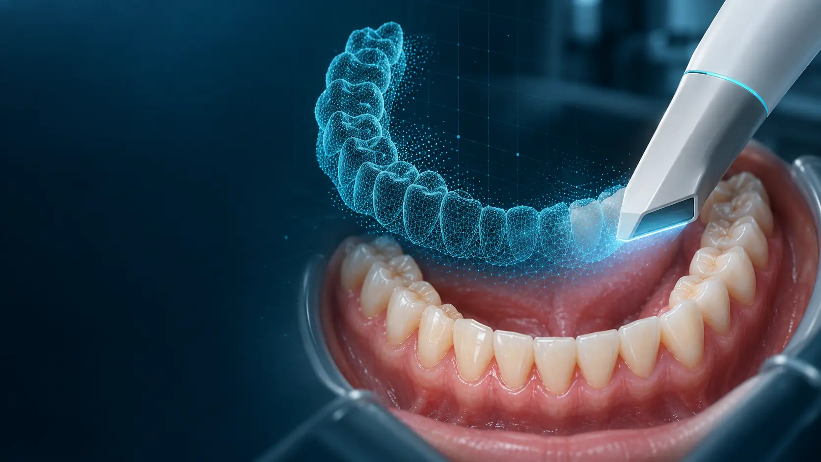
What is an intraoral scan?
Many optical images become a precise 3D surface model.
The scanner is guided across the dental arches. Cameras and light continuously record the visible surfaces while software assembles the information into a spatial model in real time. No X-ray radiation is produced.
What does it capture?
Teeth, preparation margins, gum surfaces and the bite.
The scan shows visible structures in great detail. Cone beam CT (CBCT) shows the bone, nerves and maxillary sinus beneath the surface. Together, the two datasets provide the full basis for digital planning.
Why does it matter for implants?
The future tooth guides the planning of the implant.
We combine the surface scan with the CBCT scan and the planned crown or bridge. This lets us plan the implant position around the available bone, safety margins, bite, appearance and access for cleaning.
What does the patient experience?
No conventional impression tray where the treatment step can be completed digitally.
Scanning can be performed in sections, paused and selectively supplemented. The finished model can be reviewed together immediately, making findings, planning and the future restoration easier to understand.