
3D diagnostics
3D diagnostics at ZAK
See more.
Plan with purpose.
At ZAK, no implant is planned without prior DVT diagnostics. Digital volume tomography creates a three-dimensional dataset of the jaw region and reveals spatial relationships that cannot be represented completely in a two-dimensional X-ray.
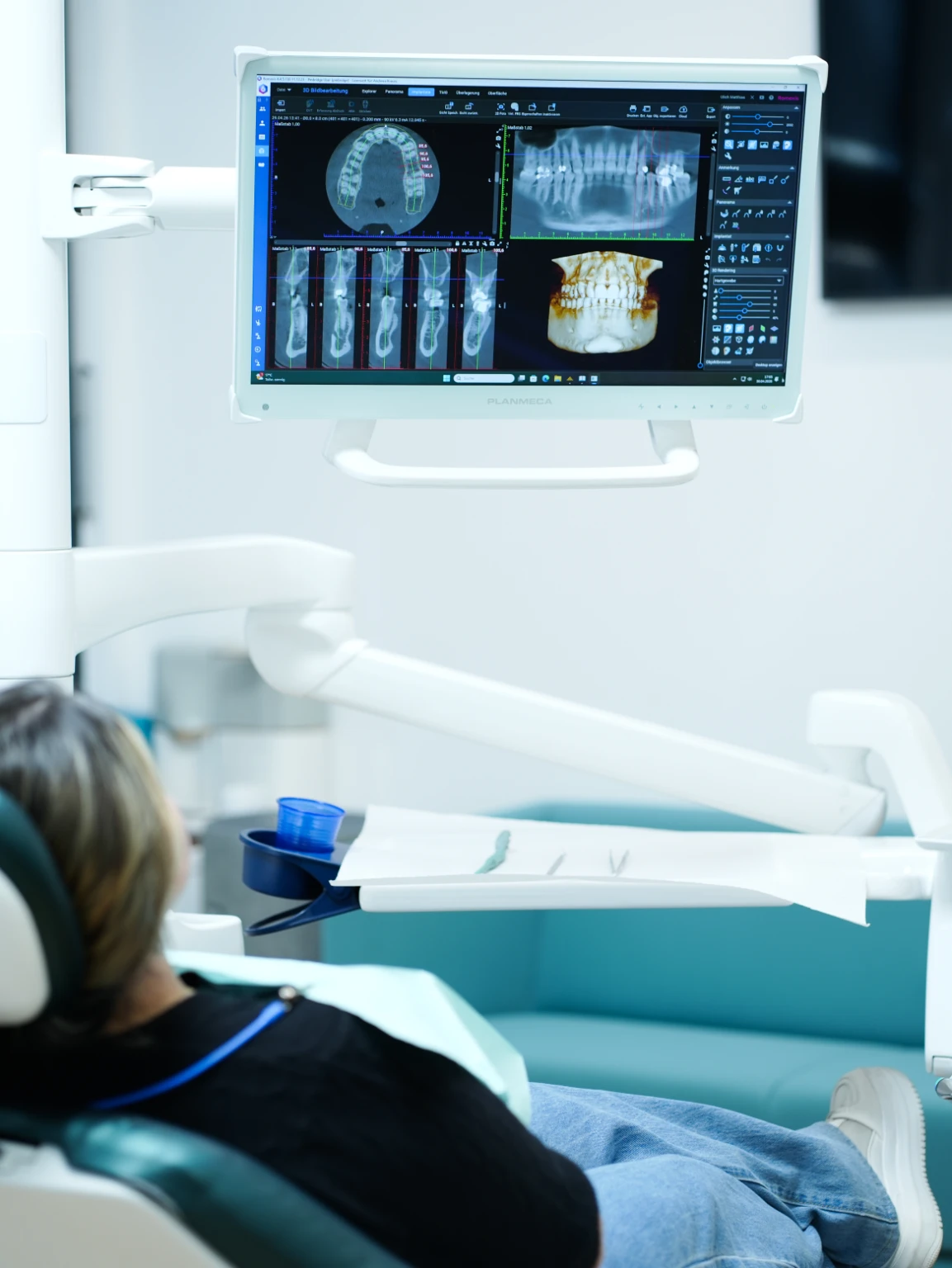
Spatial assessment
What a DVT scan can show
For implant planning, the height, width and shape of the available bone and the position of sensitive anatomical structures can be decisive. Images can be viewed in freely selectable sectional planes and as a spatial reconstruction.
- Bone: three-dimensional volume and anatomical shape at the intended implant site
- Safety distances: relation to the nerve canal, maxillary sinus and neighbouring tooth roots
- Planning basis: virtual preparation of implant position, length and diameter
- Complex situations: additional orientation for bone loss, augmentation, sinus lift or anatomical variations
Digital · Ultra Low Dose
3D information with modern dose-reduction technology.
Ultra Low Dose certified
ZAK's radiography is fully digital and uses Ultra Low Dose certified technology. It enables three-dimensional imaging with dose-optimised protocols selected for the diagnostic task.
Individually configured for every implant
DVT is a standard part of every implant plan at ZAK. The field of view, resolution and protocol are nevertheless selected for the specific region and question, providing the exact dataset required for spatial planning.
The field of view and medical indication are determined after an individual examination. Radiation protection is guided, among other sources, by the German S2k guideline on dental digital volume tomography (AWMF 083-005).
Digital implant planning
Anatomy and the final restoration are planned together.
CBCT provides the three-dimensional anatomy. The prosthetic goal defines where the future tooth should stand in terms of function, aesthetics and hygiene. Together, these elements form a plan that connects the available bone with the final restoration.
- 01CBCT
Three-dimensional assessment of bone, nerve pathways, the maxillary sinus and neighbouring structures.
- 02Prosthetic goal
The position, shape, function and cleanability of the future crown or bridge are defined in advance.
- 03Virtual planning
Implant position and dimensions are coordinated between anatomical limits and the planned restoration.
- 04Surgical transfer
The digital plan is implemented under clinical control, using a surgical guide when indicated.